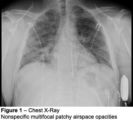
tree in bud opacities in lungs
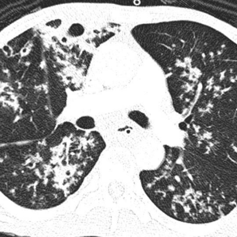
Opportunistic lung infection in a 73-year-old female patient with selective IgG3 deficit. Tree-in-bud opacities appear as tiny centrilobular branching structures on CT most often in the lung periphery which resemble budding trees Figure 18-4.

Tree In Bud Pattern Pulmonary Tb Eurorad
In radiology the tree-in-bud sign is a finding on a CT scan that indicates some degree of airway obstruction.
. We here describe an unusual cause of TIB during the COVID-19 pandemic. The tree-in-bud sign is a nonspecific imaging finding that implies impaction within bronchioles the smallest airway passages in the lung. A young male patient who had a history of fever cough and respiratory distress presented in the emergency department.
Multiple causes for tree-in-bud TIB opacities have been reported. TIB opacities are also associated with bronchiectasis and small airways obliteration resulting in mosaic air trapping. Citation DOI article data.
Usually somewhat nodular in appearance the tree-in-bud pattern is generally most pronounced in the lung periphery. Respiratory infections 72 with TB. Multiple causes for tree-in-bud TIB opacities have been reported.
Distal pulmonary vasculature More specifically the pattern can be manifest because of the following disease processes often in combination. Simply put the tree-in-bud pattern can be seen with two main sites of disease 3. The tree-in-bud pattern is commonly seen at thin-section computed tomography CT of the lungs.
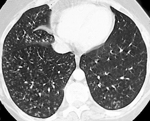
Bronchial disorders CT lung. It consists of small centrilobular nodules of soft-tissue attenuation connected to multiple branching linear structures of similar caliber that originate from a single stalk. Tree-in-bud sign or pattern describes the CT appearance of multiple areas of centrilobular nodules with a linear branching pattern.
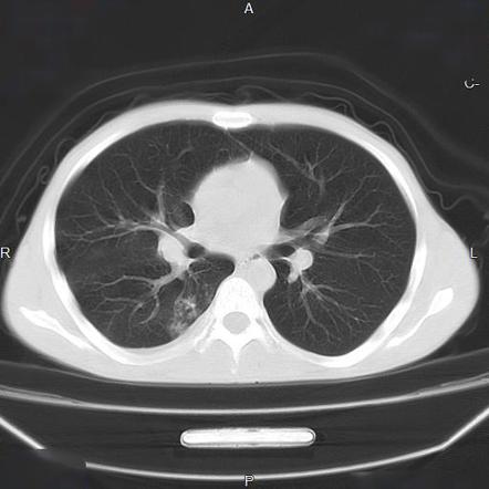
Originally reported in cases of endobronchial spread of Mycobacterium tuberculosis this pattern is now. There are widespread TIB opacities throughout both lungs with predominance in the anterior nondependent portions of the lungs a finding that would normally suggest a diagnosis other than aspiration. However to our knowledge the relative frequencies of the causes have not been evaluated.
The purpose of this study was to determine the relative frequency of causes of TIB opacities and identify patterns of disease associated with TIB opacities. Although initially described in patients with endobronchial tuberculosis it is now recognised in a large number of conditions. Bronchiolesfilled with pus or inflammator.
1 refers to a pattern seen on thin-section chest CT in which centrilobular bronchial dilatation and filling by mucus pus or fluid resembles a budding tree Fig. Multiple causes for tree-in-bud TIB opacities an imaging pattern usually seen on chest CT have been reported. Distal airways more common 2.
These are due to filling of the distal bronchioles and involvement of the adjacent alveoli most often caused by infectious bronchiolitis bronchitis and aspiration. In addition the centrilobular nodules have a branching configuration and appear to arise from a stalk otherwise known as a tree-in-bud pattern. Tree-in-bud TIB appearance in computed tomography CT chest is most commonly a manifestation of infection.
These nodules are centered within the secondary pulmonary lobule without involvement of the subpleural lung compatible with a centrilobular distribution. Usually somewhat nodular in appearance the tree-in-bud pattern is generally most pronounced in the lung periphery. However in any individual case all causes of.
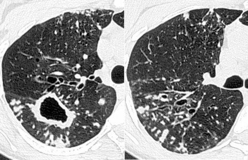
The differential for this finding includes malignant and inflammatory etiologies either infectious or sterile. Bronchial disorders CT lung. HRCT image on the axial plane depicts bronchiectasis associated with peribronchial alveolar consolidation in the middle lobe and to a less extent in the lingula.
Ad Get the Latest On The First Signs of Lung Cancer In This Article. 25k views Answered 2 years ago. However to our knowledge the relative frequencies of the causes have not been evaluated.
1 refers to a pattern seen on thin-section chest CT in which centrilobular bronchial dilatation and filling by mucus pus or fluid resembles a budding tree Fig. Concomitant tree-in-bud opacities in the lower lobes are also depicted. 11 TIB opacities represent a central imag- Background.
Tree-in-bud TIB opacities are a common imaging finding on thoracic CT scan.
2

Tree In Bud Pattern Pulmonary Tb Eurorad
Tree In Bud Sign Lung Radiology Reference Article Radiopaedia Org

Co Rads 2 With Tree In Bud Sign A 27 Year Old Male Attended The Download Scientific Diagram
Tree In Bud Sign Lungs
View Of Tree In Bud The Southwest Respiratory And Critical Care Chronicles
Tree In Bud Sign Lungs

Tree In Bud Sign Lung Radiology Reference Article Radiopaedia Org
Tree In Bud Pattern Pulmonary Tb Eurorad
2

Pdf Tree In Bud
View Of Tree In Bud The Southwest Respiratory And Critical Care Chronicles

Scielo Brasil Tree In Bud Pattern Tree In Bud Pattern

Tree In Bud Sign And Bronchiectasis Radiology Case Radiopaedia Org
2

Tree In Bud Sign Lung Radiology Reference Article Radiopaedia Org

Ct Scan Of Chest Revealing Scattered Tree In Bud Opacities In Both Download Scientific Diagram
It Is Not Always Tuberculosis Tree In Bud Opacities Leading To A Diagnosis Of Sarcoid Shm Abstracts Society Of Hospital Medicine

Pdf Tree In Bud Semantic Scholar